Introduction to Tissues: The Building Blocks of Life
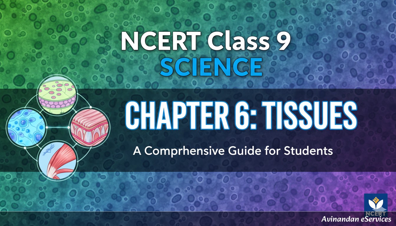
Welcome, students, to a deep dive into Chapter 6 of your Class 9 Science curriculum: Tissues. In the previous chapter, we explored the fascinating world of the cell, discovering it as the fundamental structural and functional unit of all living organisms. We learned that single-celled organisms, like Amoeba, perform all life functions—movement, intake of food, gaseous exchange, and excretion—within that single cell. But what about multicellular organisms like plants, animals, and even ourselves? How do these complex beings manage millions of cells to work in harmony? The answer lies in the concept of tissues.
In multicellular organisms, cells group together to perform specific functions. This organized clustering of cells is what we call a tissue. A tissue is formally defined as a group of cells that are similar in structure and/or work together to achieve a particular function. For example, in our bodies, muscle cells contract and relax to cause movement, and nerve cells carry messages. In plants, vascular tissues conduct water and food from one part of the plant to another. This grouping allows for a division of labour. Just like in a large factory where different departments handle specific tasks to improve overall efficiency, tissues in a multicellular organism allow for specialized cells to focus on a single function, leading to a much more efficient and organized system. This chapter will unravel the different types of tissues found in plants and animals, exploring their unique structures and the vital roles they play.
Are Plants and Animals Made of the Same Types of Tissues?
Before we classify the various tissues, a fundamental question arises: are the tissues in plants and animals similar? A brief comparison of their lifestyles and functions gives us a clear answer: no. Plants and animals have evolved under very different pressures and for different modes of existence, leading to significant differences in their tissue organization.
- Mobility: The most obvious difference is movement. Animals are motile; they move around in search of food, mates, and shelter. This requires a high amount of energy. Consequently, most animal tissues are living and have high energy demands. Plants, on the other hand, are stationary or fixed. They don't need to move, so their energy needs are lower. A large proportion of plant tissues, like sclerenchyma and xylem, are supportive and composed of dead cells, which provide mechanical strength without consuming energy.
- Growth Patterns: Growth in animals is generally uniform and stops after reaching a certain age or size. The entire body grows more or less simultaneously. In plants, growth is not uniform; it is restricted to specific regions called meristems. These regions contain actively dividing cells throughout the plant's life, allowing for continuous growth in length and girth.
- Structural Requirements: Plants need to stand upright against gravity and withstand environmental stresses like wind. This requires strong, rigid, supportive tissues. Animals, with their active locomotion, need tissues that allow for flexibility, movement, and rapid communication, such as muscles and nervous tissue, which are far more complex and developed than in plants.
These fundamental differences in lifestyle and function have led to the evolution of distinct types of tissues in plants and animals, which we will now explore separately.
Plant Tissues: The Foundation of Flora
Plant tissues can be broadly categorized into two main types based on their dividing capacity: Meristematic Tissues and Permanent Tissues.
Meristematic Tissue: The Growth Engine
Imagine the very tip of a growing shoot or a root pushing its way through the soil. The reason for this growth is meristematic tissue. These tissues are composed of actively dividing cells and are found in the growing regions of the plant. They are the plant's engine of growth, responsible for increasing its length and diameter.
Characteristics of Meristematic Cells:
- Actively Dividing: Their primary function is cell division (mitosis) to produce new cells.
- Dense Cytoplasm: They are filled with dense cytoplasm and lack large central vacuoles, as vacuoles are primarily for storage and providing turgidity, which are functions of mature cells.
- Thin Cellulose Walls: The cell walls are thin and elastic to allow for easy cell division and growth.
- Prominent Nuclei: They possess large, conspicuous nuclei that control the high metabolic activity required for cell division.
Types of Meristematic Tissues
Based on their location in the plant body, meristematic tissues are classified into three types:
- Apical Meristem: Found at the growing tips (apices) of stems and roots. Its primary role is to increase the length of the plant. This is known as primary growth. When you see a plant growing taller, it's the work of the apical meristem.
- Lateral Meristem (Cambium): Located on the sides or lateral regions of the stem and root. It is responsible for the increase in the girth or diameter of the plant. This is known as secondary growth, which makes the stem and root thicker and stronger over time.
- Intercalary Meristem: Situated at the base of the leaves or internodes (the part of the stem between two nodes). It helps in the longitudinal growth of organs like leaves and is particularly common in grasses. It allows for the regrowth of parts eaten by grazing animals.
Permanent Tissue: Specialised for Function
What happens to the new cells produced by meristematic tissues? They grow, mature, and gradually lose their ability to divide. They take up a specific role and form a permanent tissue. This process, where cells take on a permanent shape, size, and function, is called differentiation. Permanent tissues are classified into two types: Simple Permanent Tissues and Complex Permanent Tissues.
Simple Permanent Tissues
These tissues are 'simple' because they are composed of only one type of cell, all performing the same function.
- Parenchyma: This is the most common and unspecialised simple permanent tissue. Its cells are living, with thin cell walls and typically have large intercellular spaces between them. Its functions are diverse: it primarily serves as a packing tissue and provides support, but its main role is food storage. When it contains chlorophyll and performs photosynthesis, it is called chlorenchyma (found in leaves). In aquatic plants, parenchyma cells have large air cavities to provide buoyancy, and this type is called aerenchyma.
- Collenchyma: This tissue provides flexibility to plant parts like the stem and leaf stalks (petioles), allowing them to bend without breaking. You can see its effect when you bend a young stem and it doesn't snap. The cells are living, elongated, and are irregularly thickened at the corners due to the deposition of pectin. They have very little intercellular space.
- Sclerenchyma: This is the tissue that makes plants hard and stiff. Think of the husk of a coconut—it's made of sclerenchymatous tissue. The cells of this tissue are dead. They have very thick, lignified walls (lignin is a chemical substance that acts like cement and hardens the cell wall). There are no intercellular spaces. Its sole purpose is to provide mechanical strength and protection to the plant. It exists in two forms: long, narrow fibres and irregularly shaped sclereids (found in the gritty pulp of fruits like pears and guavas).
Complex Permanent Tissues
Complex tissues are 'complex' because they are made of more than one type of cell. All these different cells coordinate to perform a common function. The two types of complex tissues in plants are xylem and phloem. Together, they form the vascular bundle, which is the transport system of the plant.
- Xylem: Xylem is the water-conducting tissue. It transports water and dissolved minerals absorbed by the roots upwards to the rest of the plant. It consists of four different types of cells:
- Tracheids and Vessels: These are elongated, tube-like structures with thick, lignified walls. They form a continuous pipeline for efficient water transport. Both are dead cells at maturity.
- Xylem Parenchyma: These are the only living cells in the xylem. They store food and help in the lateral conduction of water.
- Xylem Fibres: These are sclerenchymatous fibres that are mainly supportive in function.
- Phloem: Phloem is the food-conducting tissue. It transports food, prepared in the leaves through photosynthesis, to other parts of the plant. This process is called translocation. Unlike xylem, phloem can transport materials in both upward and downward directions. It is composed of four types of elements:
- Sieve Tubes: These are tube-like structures with perforated end walls called sieve plates. They are the main conducting channels for food. Mature sieve tubes lack a nucleus.
- Companion Cells: These cells are closely associated with sieve tubes. They have a nucleus and other organelles, and they regulate the functions of the sieve tubes.
- Phloem Parenchyma: These cells store food and help in transport.
- Phloem Fibres: These are sclerenchymatous fibres that provide mechanical strength. They are the only dead cells in the phloem.
Protective Tissue in Plants
Plants also have tissues dedicated to protection from the \texternal environment.
- Epidermis: This is the outermost single layer of cells covering the entire plant. It is like the plant's skin. Its main function is protection. The cells are flat and have no intercellular spaces. In dry habitats, the epidermis is often thicker and may be covered by a waxy, water-resistant layer called the cuticle (made of cutin), which prevents water loss. The epidermis of the leaf has small pores called stomata. Each stoma is guarded by two kidney-shaped cells called guard cells, which regulate the opening and closing of the pore for gaseous exchange and transpiration.
- Cork: As a plant grows older, the outer protective epidermis on the stem is replaced by a secondary meristem that forms a multi-layered, tough outer layer called cork or bark. The cells of cork are dead and compactly arranged without intercellular spaces. They have a chemical called suberin deposited in their walls, which makes them impermeable to gases and water, providing excellent protection against mechanical injury, temperature \textremes, and pathogens.
Animal Tissues: The Fabric of Fauna
Animal bodies are complex machines that require highly specialized tissues for their diverse functions like movement, response to stimuli, digestion, and circulation. On the basis of their functions, animal tissues are broadly classified into four main types: Epithelial Tissue, Connective Tissue, Muscular Tissue, and Nervous Tissue.
Epithelial Tissue: The Covering and Protective Layer
Epithelial tissue is the covering or lining tissue of the body. It covers most organs and cavities within the body and also forms a barrier to keep different body systems separate. The skin, the lining of the mouth, the lining of blood vessels, and lung alveoli are all made of epithelial tissue. The cells are tightly packed and form a continuous sheet with almost no intercellular spaces. They are separated from the underlying tissue by an \textracellular fibrous basement membrane.
Types of Epithelial Tissues
Epithelial tissues are classified based on the shape and arrangement of their cells.
- Simple Squamous Epithelium: Composed of a single layer of \textremely thin, flat cells, resembling tiles on a floor. Due to their thinness, they are selectively permeable and are involved in filtration and diffusion. They are found lining blood vessels (where they are called endothelium) and lung alveoli.
- Stratified Squamous Epithelium: This tissue consists of cells arranged in multiple layers (strata). This layered arrangement prevents wear and tear. Our skin, which is constantly subjected to friction, is a prime example of stratified squamous epithelium.
- Cuboidal Epithelium: As the name suggests, this tissue is composed of a single layer of cube-shaped cells. It is commonly found in the lining of kidney tubules and ducts of salivary glands, where it provides mechanical support and is involved in secretion and absorption.
- Columnar Epithelium: Composed of a single layer of tall, pillar-like cells. It is found where absorption and secretion occur, such as the inner lining of the intestine. In the respiratory tract, these cells often have hair-like projections called cilia on their outer surfaces. This ciliated columnar epithelium helps to move mucus and other substances along the tract.
- Glandular Epithelium: Sometimes, a portion of the epithelial tissue folds inward to form a multicellular gland that can secrete substances. This is known as glandular epithelium, found in sweat glands, tear glands, and salivary glands.
Connective Tissue: The Binding and Supporting Matrix
Connective tissue is the most abundant and widely distributed tissue in the animal body. Its primary function is to link, support, and connect other tissues or organs. The cells of connective tissue are loosely spaced and are embedded in an intercellular matrix. This matrix can be jelly-like, fluid, dense, or rigid, and its nature determines the function of the tissue.
Types of Connective Tissues
- Blood: This is a fluid connective tissue. The fluid matrix is called plasma, in which red blood cells (RBCs), white blood cells (WBCs), and platelets are suspended. Plasma transports food, hormones, and waste materials. RBCs transport oxygen, WBCs fight diseases, and platelets help in blood clotting.
- Bone: This is a rigid connective tissue that forms the body's framework or skeleton. Bone cells (osteocytes) are embedded in a hard matrix composed of calcium and phosphorus compounds. Bone anchors muscles, protects vital organs, and supports the body.
- Cartilage: This is a firm but flexible connective tissue. It has widely spaced cells embedded in a solid matrix of proteins and sugars. It smoothens bone surfaces at joints and is also present in the nose, ear, trachea, and larynx.
- Ligaments and Tendons: These are types of fibrous connective tissue.
- Ligaments are very elastic and connect one bone to another, holding joints together.
- Tendons are strong and have limited flexibility. They connect skeletal muscles to bones.
- Areolar Tissue: This is a loose connective tissue found between the skin and muscles, around blood vessels and nerves, and in the bone marrow. It fills the space inside organs, supports internal organs, and helps in the repair of tissues.
- Adipose Tissue: This tissue is specialized for fat storage. It is found below the skin and between internal organs. The cells of this tissue (adipocytes) are filled with fat globules. It acts as an insulator, protecting the body from cold, and also serves as a shock-absorbing cushion.
Muscular Tissue: The Engine of Movement
All movement in the body is brought about by the contraction and relaxation of muscular tissue. The cells of this tissue are elongated and are also called muscle fibres. They contain special contractile proteins that enable movement.
Types of Muscular Tissues
Based on their structure, location, and function, we can differentiate three types of muscle tissues.
- Striated Muscles (Skeletal or Voluntary Muscles): These muscles are attached to bones and are responsible for body movements like walking, running, and lifting. We can move them according to our will, hence they are called voluntary muscles. The cells are long, cylindrical, unbranched, and multinucleate (having many nuclei). They show alternate light and dark bands or striations under a microscope.
- Smooth Muscles (Unstriated or Involuntary Muscles): These muscles are found in the walls of internal organs such as the alimentary canal, blood vessels, and iris of the eye. Their movement is not under our conscious control, hence they are called involuntary muscles. The cells are long with pointed ends (spindle-shaped) and are uninucleate (having a single nucleus). They do not show any striations.
- Cardiac Muscles (Heart Muscles): These muscles are found only in the walls of the heart. They are involuntary in action. Structurally, they are a hybrid of striated and smooth muscles. The cells are cylindrical, branched, and uninucleate. They show faint striations. Cardiac muscles contract and relax rhythmically and tirelessly throughout a lifetime to pump blood.
Nervous Tissue: The Communication Network
Nervous tissue is highly specialized for being stimulated and then transmitting that stimulus very rapidly from one place to another within the body. The brain, spinal cord, and all the nerves in our body are composed of nervous tissue. This tissue is responsible for control and coordination.
The cells of the nervous tissue are called nerve cells or neurons. A neuron consists of three main parts:
- Cell Body (or Cyton): This contains a nucleus and cytoplasm, representing the main metabolic center of the cell.
- Dendrites: These are short, branched, hair-like \textensions arising from the cell body. They receive nerve impulses from other neurons.
- Axon: This is a single, long, cylindrical process that \textends from the cell body. It is responsible for transmitting nerve impulses away from the cell body to the next neuron or to a muscle/gland. The axon may be covered by a protective sheath called the myelin sheath.
The combination of a neuron's axon and its protective sheaths is called a nerve fibre. Many nerve fibres bound together by connective tissue make up a nerve, forming the body's intricate communication network.
Important Questions and Answers (from NCERT Exercise)
Q1: Define the term "tissue".
Answer: A tissue is a group of cells that are similar in structure and are organized together to perform a specific function. This organization allows for a division of labour in multicellular organisms, leading to increased efficiency.
Q2: Name the types of simple tissues in plants.
Answer: The three types of simple permanent tissues in plants are:
- Parenchyma: A living tissue with thin walls, used for storage and as packing tissue.
- Collenchyma: A living tissue with unevenly thickened corners, providing flexibility and mechanical support.
- Sclerenchyma: A dead tissue with thick, lignified walls, providing hardness and mechanical strength.
Q3: What is the specific function of the cardiac muscle?
Answer: The specific function of the cardiac muscle is to contract and relax rhythmically and continuously throughout a person's life. This rhythmic pumping action of the heart, which is involuntary, is responsible for circulating blood throughout the body, supplying oxygen and nutrients and removing waste products.
Q4: Differentiate between striated, unstriated and cardiac muscles on the basis of their structure and site/location in the body.
Answer:
| Feature | Striated Muscles | Unstriated (Smooth) Muscles | Cardiac Muscles |
|---|---|---|---|
| Cell Shape | Long, cylindrical, unbranched | Long, spindle-shaped (pointed ends) | Cylindrical, branched |
| Nucleus | Multinucleate (many nuclei), peripheral | Uninucleate (single nucleus), central | Uninucleate (single nucleus), central |
| Striations | Present (alternate light and dark bands) | Absent | Present (faint striations) |
| Location/Site | Attached to bones (e.g., in limbs, trunk, face) | Walls of internal organs (e.g., stomach, intestine, blood vessels, iris) | Walls of the heart |
| Action | Voluntary (under conscious control) | Involuntary (not under conscious control) | Involuntary (not under conscious control) |
Q5: Name the tissue responsible for movement in our body.
Answer: The tissue responsible for movement in our body is Muscular Tissue. Its cells, also called muscle fibres, have the ability to contract and relax, which generates force and produces movement of various body parts.
Chapter Summary
Here are the key takeaways from our comprehensive study of tissues:
- A tissue is a group of similar cells performing a specific function.
- Plant and animal tissues differ significantly due to their different modes of life (sedentary vs. mobile).
- Plant tissues are of two main types: Meristematic (dividing) and Permanent (differentiated).
- Meristematic tissue is found in growing regions and is of three types: apical, lateral, and intercalary.
- Permanent tissues are of two types: Simple (Parenchyma, Collenchyma, Sclerenchyma) and Complex (Xylem, Phloem).
- Xylem transports water and minerals, while Phloem transports food.
- Animal tissues are classified into four types: Epithelial, Connective, Muscular, and Nervous.
- Epithelial tissue is a protective covering and lining, with types like squamous, cuboidal, and columnar.
- Connective tissue binds and supports, and includes blood, bone, cartilage, ligaments, tendons, areolar, and adipose tissues.
- Muscular tissue is responsible for movement and includes striated (voluntary), smooth (involuntary), and cardiac (involuntary) muscles.
- Nervous tissue, made of neurons, is responsible for transmitting stimuli and coordinating bodily functions.